After a string of recent posts all shanti-shanti and fluffy, let’s get back to some hardcore science.
Vision science is an amazing field. Truly, retail optometry doesn’t do any semblance of justice to the depth and breadth of research being done on the subject of human (and animal) vision. The sheer quantity of data and analysis available to you right now, today, is a testimony to human curiosity and ingenuity.
One of the eyeball topics you might find interesting is contrast adaptation.
It’s particularly interesting because it’s a) not entirely understood, b) heavily being researched and c) has curious implications on the brain’s role in vision and the development of myopia.
Before we lose the not-geeks … I use contrast adaptation based techniques in the (paid) program quite a bit. Since the program is regular-human friendly I don’t deep dive into the science there. The program is strictly about improving your eyesight, the quickest and least involved way possible. But if you dig some of the (very large) back story, articles like this one might be up your alley. Enjoy!
If you’re still new to the “how to find relevant science”, here’s a tip for you:
Always start with looking for animal studies.
Since us humans have little respect for life that isn’t our species, the more adventurous and less carefully worded analysis is always found in animal studies.
There’s always a lot of tip-toeing when it comes to drawing conclusions on human eyesight studies. Must not offend the sponsors and those who hold dominion over it all. (lens manufacturers) So it’s much harder to get a good footing if you start with the human science.
So you start with animal studies, and then use the keywords there to find related human studies.
Tricky. But absolutely fun sleuthing.
Here’s an example of a chicken study that talks about contrast sensitivity:
![]() Changes in contrast sensitivity induced by defocus and their possible relations to emmetropization in the chicken.
Changes in contrast sensitivity induced by defocus and their possible relations to emmetropization in the chicken.
Source: http://europepmc.org/abstract/med/11687557
PURPOSE: To test whether the level of contrast adaptation (CA) relates to refractive development in the chicken. (CA refers to a spatial frequency-selective increase of suprathreshold contrast sensitivity after exposure to low-contrast patterns).
METHODS: CA was determined in individual chicks by comparing their optomotor gain in response to drifting low-contrast stripe patterns before and after treatment with spectacle lenses. The amount of CA was compared with the loss of contrast predicted from defocus at the tested spatial frequency. The reversion of CA and recovery from deprivation myopia were studied while the retinal image features were controlled by forcing the animals to watch spatially filtered digital video clips.
RESULTS: CA was induced by wearing positive and negative lenses for 1.5 hours, both without and with cycloplegia, but was less pronounced in the case of positive lenses when accommodation was intact. The amount of CA at a tested spatial frequency was predicted from the loss of contrast calculated from the modulation transfer function for a defocused optical system. Watching low-pass-filtered video clips induced deprivation myopia and inhibited recovery from it. It also prevented the reversal of CA that was previously induced by deprivation. Both recovery from deprivation myopia and recovery from CA occurred with sharp video clips, although less so than with normal visual exposure.
CONCLUSIONS: CA changes with retinal image sharpness and occurs even when accommodation is intact. Because CA correlates with myopia induced by frosted occluders, negative lenses, and low-pass-filtered video clips, and its reversal correlates with recovery from myopia, it is possible that shifts in CA may represent a signal related to refractive error development.”
—
This is the clue hunting, when it comes to understanding myopia. Lots of words here that might be new, do Google about if you’re new to the topic at large. (or read back through the blog where we cover a lot of these topics)
From the chickens, you might search for contrast adaptation studies in human. Lots of those, but not quite as many dealing with retinal contrast adaptation, unfortunately.
Contrast adaptation in human retina and cortex.
Source: http://www.ncbi.nlm.nih.gov/pubmed/11581221
PURPOSE:
Although cortical contrast adaptation has been extensively studied with both psychophysical and electrophysiological techniques, little is known about retinal contrast adaptation in humans.
METHODS:
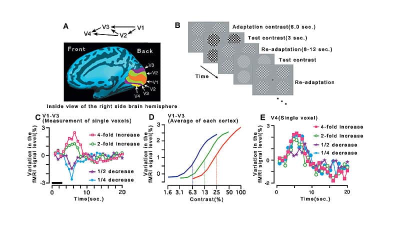
Retinal and cortical long-term contrast adaptation was assessed with simultaneous measurement of pattern electroretinogram (PERG) and cortical visual evoked potentials (VEPs). This study involved three approaches: sampling of the contrast transfer function from 2.7% to 98% with adaptation to high (98%) and low (7.3%) contrasts, linearity of adaptation effects, and transfer of contrast adaptation between parallel and orthogonal grating orientations.
RESULTS:
Contrast adaptation affected retinal and cortical recordings quite differently. The VEP showed a sigmoid contrast transfer function, which was shifted toward higher contrasts (by a factor of 1.9), whereas amplitudes at higher test contrasts were enhanced to 127%. The PERG decreased in amplitude to approximately 90%, and the latency was significantly reduced by 4 to 6 msec (P < 0.05). All measured effects were linear with adaptation contrast. Orientation played no role in the PERG results, whereas the VEP was enhanced to 125% when tested parallel and to 150% when tested orthogonal to adaptation.
CONCLUSIONS:
VEP results confirm and extend previous findings and fit well with single-cell recordings. The PERG findings suggest that retinal contrast adaptation occurs and mainly operates in the temporal domain, comparable to rapid gain-control findings in cats and primates.
—
So we have chicken science, and we have interesting findings on eye and brain long-term contrast adaptation. We go from animals, to some relatively abstract human science. Depending on your levels of curiosity, further searches on scholar.google.com can lead you deeper into the rabbit hole.
 Near work-induced contrast adaptation in emmetropic and myopic children.
Source: http://www.ncbi.nlm.nih.gov/pubmed/22562511
PURPOSE:
Contrast adaptation may induce an error signal for emmetropization. This research aims to determine whether reading causes contrast adaptation in children and, if so, to determine whether myopes exhibit greater contrast adaptation than emmetropes.
METHODS:
Baseline contrast sensitivity was determined in 34 emmetropic and 34 spectacle-corrected myopic children for 0.5, 1.2, 2.7, 4.4, and 6.2 cycles per degree (cpd) horizontal sine-wave gratings. Effects of near tasks on contrast sensitivity were determined during periods spent looking at a 6.2 cpd horizontal grating and during periods spent reading lines of English text, with 1.2 cpd row frequency and 6 cpd stroke frequency.
RESULTS:
Both emmetropic and myopic groups (mean ± SD; age, 10.3 ± 1.4 years) showed reduced contrast sensitivity during both near tasks, with greatest overall adaptation at 6.2 cpd. Adaptation induced by viewing the grating (0.15 ± 0.17 log unit
CONCLUSIONS:
Grating and reading tasks induced contrast adaptation; viewing horizontal gratings induced greater adaptation than reading, and myopes exhibited greater adaptation than emmetropes. Contrast adaptation effects may underlie findings of prolonged near work being associated with myopia. However, our research does not show whether this is consequential or causal.
—
And that’s where things continue to get more interesting. Notice that trend I mentioned earlier, on how conclusions become worded much more carefully, anytime we’re looking at findings that may potentially be damning to the retail vision world, and related deep pockets.
It also pays off to go wide on research. These guys don’t necessarily talk to each other, read each other’s research, and there are always politics involved in one group over another. The only way to get a full picture is by looking at a number of studies and articles on the subject and look for common elements and findings.
 Blur Adaptation and Myopia
Source: http://journals.lww.com/optvissci/Abstract/2004/07000/Blur_Adaptation_and_Myopia.16.aspx
Previous studies have demonstrated a significant improvement in visual resolution during sustained periods of retinal defocus. This appears to result from perceptual adaptation designed to restore the perceived contrast of the degraded image. However, it is unclear whether perceptual adaptation to sustained blur is present in all individuals or only in certain subgroups, such as those who have been chronically exposed to sustained periods of blur due to uncorrected ametropia. Accordingly, the present study examined the effects of sustained retinal defocus on both high-and low-contrast visual acuity in emmetropes (n = 13) and myopes (n = 18). Subjects were required to view through +2.50-D spherical lenses worn over their distance refractive correction for a continuous 2-hour period. A significant improvement in both Landolt C and grating visual acuity measured through the fogging lenses was observed in both refractive groups. Although the mean change in grating visual acuity was significantly greater for the myopic subjects, the improvements in Landolt C acuity observed in the emmetropes and myopes were statistically equivalent. We hypothesize that the improvement in visual acuity results from perceptual adaptation to the blurred images, which may occur at central sites within the visual cortex.
—
That’s some hard hitting stuff, right there.
Remember when I talk about how vision happens in your brain? 90% of what you read and specific suggestions I make, all seem trivial and simple. You know why?
Because I know my audience. (the majority of which isn’t you, still reading this article here)
Most people want to know that a) I’m the real deal and that b) they are reasonably going to get the outcome they’re looking for. (better eyesight) Past that, 90% of people have zero interest in digging through vision science papers, most of which are filled with data that one might reasonably consider to be ego stroking and purposeful adding of complexity to inflate the perceived importance of the study.
So when you read most of my things, keep that in mind. I do casually mention the decade of research, though the final actionable pieces are all designed to be conducive to habit building, and not putting people to sleep while reading. ;-)
How about that last excerpt article though? Damn.
If you really want to dive in, you might go for reading further into this one. (though, not exactly necessary for your intended purpose of improving your eyesight)
![]() How do attention and adaptation affect contrast sensitivity?
How do attention and adaptation affect contrast sensitivity?
Source: http://jov.arvojournals.org/article.aspx?articleid=2193083
The visual environment is rich in information. Despite the fact that the brain uses almost a quarter of the body’s metabolic resources, the energy available at each instant is insufficient to process all incoming sensory information (Lennie, 2003). The brain has to maximize performance while maintaining energy expenditures within its metabolic limits to attain our astonishing perceptual abilities. Several mechanisms allocate perceptual resources to optimize our sensitivity to match salient parts of visual scenes. Spatial attention and contrast adaptation are two such managing mechanisms. In this study, we investigated whether and how the adaptation state and the attentional effects on contrast sensitivity interact. In particular, we explored whether attention can overcome adaptation and restore contrast sensitivity.

