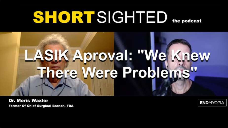
Your darling old dubiously bearded eye guru finally got around to asking our most favorite former FDA connect, Dr. Morris Waxler, to talk to us about LASIK.
It was even more of a hair raising conversation than I expected.
A solid hour of him really laying into all the potentially serious and unfixable consequences of the procedure, as well as the lack of approval requirements for medical side effects, and the “cowboy” attitudes of surgeons. It’s one thing to read a transcript or see statistics, and something entirely different to hear the former Chief Of Surgical Branch at the FDA to put it in some very blunt terms.
Short Clip: They Cut The Nerves In Your Cornea
Short Clip: 30% Risk Of Permanent Dry Eyes
Short Clip: We Knew There Were Problems
Full Episode
If you ever even considered laser surgery, you owe yourself a listen to this one.
To find all episodes of the Shortsighted Podcast (normally just improvement reports), and all the platforms and apps that host us, see the podcast page.
And here is the audio of the podcast:
Below, a list of citations Dr. Waxler provided to go along with his assertions.
Citations
Chronic corneal weakness
Miyata K, Tokunaga T, Nakahara M, Ohtani S, Nejima R, Kiuchi T, Kaji Y, MD, Oshika T. Residual bed thickness and corneal forward shift after laser in situ keratomileusis J Cataract Refract Surg 2004; 30:1067–1072.
Binder PS. Analysis of ectasia after laser in situ keratomileusis: Risk factors. J Cataract Refract Surg 2007; 33:1530–1538.
Spadea L, Cantera E, Cortes M, Conocchia NE, Stewart CWM. Corneal ectasia after myopic laser in situ keratomileusis: a long-term study. Clinical Ophthalmology 2012:6 1801–1813.
Chan CCK, Hodge C, Sutton G. “External analysis of the Randleman Ectasia Risk Factor Score System: a review of 36 cases of post LASIK ectasia.” Clinical and Experimental Ophthalmology 2010; 38: 335–340.
Reinstein DZ, Waring GO III. Have You Seen the 10-year Long-term Safety Data on LASIK? Journal of Refractive Surgery Volume 22 November 2006.
Meghpara B, Nakamura H, Macsai M, Sugar J, Hidayat A, Yue BYJT, Deepak P, Edward DP. Keratectasia After Laser In Situ Keratomileusis: A Histopathologic and Immunohistochemical Study. Arch Ophthalmol . 2008;126(12):1655-1663.
Lifshitz T, Levy J, Klemperer I, Levinger S. Late Bilateral Keratectasia After LASIK in a Low Myopic Patient. J Refract Surg. 2005;21:494-496.
Reinstein DZ, Waring GO III. Have You Seen the 10-year Long-term Safety Data on LASIK? Journal of Refractive Surgery Volume 22 November 2006.
Waring GO III. A Cautionary Tale of Innovation in Refractive Surgery, ARCH OPHTHALMOL/VOL117, AUG1999 1068-1073.
Rates
Binder PS. Analysis of ectasia after laser in situ keratomileusis: Risk factors. J Cataract Refract Surg 2007; 33:1530–1538.
Spadea L, Cantera E, Cortes M, Conocchia NE, Stewart CWM. Corneal ectasia after myopic laser in situ keratomileusis: a long-term study. Clinical Ophthalmology 2012:6 1801–1813.
Chan CCK, Hodge C, Sutton G. “External analysis of the Randleman Ectasia Risk Factor Score System: a review of 36 cases of post LASIK ectasia.” Clinical and Experimental Ophthalmology 2010; 38: 335–340.
Reinstein DZ, Waring GO III. Have You Seen the 10-year Long-term Safety Data on LASIK? Journal of Refractive Surgery Volume 22 November 2006.
Chronic pain syndrome
Rates
https://www.accessdata.fda.gov/cdrh_docs/pdf/P970053S011B.pdf Table 10
https://www.accessdata.fda.gov/cdrh_docs/pdf2/P020050S012B.pdf Table 9 Changes from preop
https://www.accessdata.fda.gov/cdrh_docs/pdf6/P060004S001B.pdf Tables 9 & 11
https://www.accessdata.fda.gov/cdrh_docs/pdf/P930016S025B.pdf Table 43
https://www.accessdata.fda.gov/cdrh_docs/pdf/P970053S009B.pdf Table 32
https://www.accessdata.fda.gov/cdrh_docs/pdf6/P060004B.pdf Table 12 Worse at 6M (mod+mark+severe)
https://www.accessdata.fda.gov/cdrh_docs/pdf/P990027S006B.pdf Table 13B
https://www.accessdata.fda.gov/cdrh_docs/pdf/P990027S004B.pdf Tables 22 & 26
Denoyer A, Landman E, Trinh L, Faure JF, Auclin F, Baudouin C. Dry Eye Disease after Refractive Surgery: Comparative Outcomes of Small Incision Lenticule Extraction versus LASIK. Ophthalmology. 2014. doi:10.1016/ j.ophtha.2014.10.004. [41 cite]
Bower KS, RK, Ryan DS, Mines MJ, and Dartt DA Chronic dry eye in PRK and LASIK: manifestations, incidence and predictive factors J Cataract Refract Surg. 2015 December ; 41(12): 2624–2634. doi:10.1016/j.jcrs.2015.06.037. [15 cited]
De Paiva CS, Chen Z, Koch DD, Hamill MB, Manuel FK, Hassan SS, et al. The incidence and risk factors for developing dry eye after myopic LASIK. Am J Ophthalmol. 2006;141(3):438–45. doi:10.1016/j.ajo.2005.10.006. [43 cites]
Shoja MR, Besharati MR. Dry eye after LASIK for myopia: Incidence and risk factors. Eur J Ophthalmol. 2007;17(1):1–6 [20 cites]
Tuisku IS, Lindbohm N, Wilson SE, Tervo TM. Dry eye and corneal sensitivity after high myopic LASIK. J Refract Surg. 2007;23(4):338–42. [ 17 cited]
Donnenfeld ED, Solomon K, Perry HD, Doshi SJ, Ehrenhaus M, Solomon R, et al. The effect of hinge position on corneal sensation and dry eye after LASIK. Ophthalmology. 2003;110(5):1023–9. discussion 9–30. doi:10.1016/ S0161-6420(03)00100-3. {33 articles cite}
Hovanesian JA, Shah SS, Maloney RK. Symptoms of dry eye and recurrent erosion syndrome after refractive surgery. J Cataract Refract Surg. 2001;27(4):577–84. [6 cited]
Albietz JM, Lenton LM, McLennan SG. Effect of laser in situ keratomileusis for hyperopia on tear film and ocular surface. J Refract Surg 2002;18:113-23 [19 cited]
Syndrome
Levitt AE, Galor A, Weiss JS, Felix ER, Martin ER, Patin DJ, Sarantopoulos KD, and Levitt RC. Chronic dry eye symptoms after LASIK parallels and lessons to be learned from other persistent post operative pain disorders. Molecular Pain 2015 11: 21 DOI 10 1186 s 12990 015 0020 7
Galor A, Zlotcavitch L, Walter SD, Felix ER, Feuer W, Martin ER, Margolis TP,, Sarantopoulos KD. Dry eye symptom severity and persistence are associated with symptoms of neuropathic pain. Br J Ophthalmol 2014;0:1–4. doi:10.1136/bjophthalmol-2014-306057.
MackieIA.Neuroparalytickeratitis.In: Fraunfelder F, Roy FH, Meyer SM, eds. Current Ocular Therapy. Philadelphia, PA: WB Saunders; 1995:452-454.
BoniniS,RamaP,OlziD,LambiaseA. Neurotrophic keratitis. Eye (Lond). 2003;17(8):989-995.
Mathers WD. The Incidence and Risk Factors for Developing Dry Eye After Myopic LASIK Procedure
January 05, 2006DOI:https://doi.org/10.1016/j.ajo.2005.11.020
Nettune GR, Pflugfelder SG. Post-LASIK Tear Dysfunction and Dysesthesia Ocul Surf. 2010 July ; 8(3): 135–145.
Shtein RM Post-LASIK dry eye. Expert Rev Ophthalmol. 2011 October ; 6(5): 575–582. doi:10.1586/eop.11.56.
Lambiase A, Sacchetti M, Mastropasqua A, Bonini S. Corneal Changes in Neurosurgically Induced Neurotrophic Keratitis. JAMA Ophthalmol. 2013;131(12):1547-1553. doi:10.1001/jamaophthalmol.2013.5064
Shaheen B, Bakir M, Jain S. Corneal Nerves in Health and Disease. Surv Ophthalmol. 2014; 59(3): 263–285. doi:10.1016/j.survophthal.2013.09.002.
Cohen E, Oriel Spierer O. Dry Eye Post-Laser-Assisted In Situ Keratomileusis: Major Review and Latest Updates. Journal of Ophthalmology,Volume 2018, Article ID 4903831, 9 pages https://doi.org/10.1155/2018/4903831
Theophanous C, Jacobs DB, and Hamrah P. Corneal Neuralgia after LASIK Optometry and Vision Science Vol 92 No 9 September 2015
Theophanous C, Jacobs DB, and Hamrah P. Corneal Neuralgia after LASIK Optometry and Vision Science Vol 92 No 9 September 2015
Rosenthal P, Baran I, Jacobs DS. Corneal pain without stain, is it real. Ocul Surf 2009: 7: 28- 40.
Chao C, Golebiowski B, Stapleton F. The role of corneal innervation in LASIK-induced neuropathic dry eye. Ocul Surf. 2014;12(1):32‐45. doi:10.1016/j.jtos.2013.09.001
Chronic vision distortions
Montés-Micó R, España E, Menezo JL.Mesopic Contrast Sensitivity Function After Laser in situ Keratomileusis J Refract Surg 2003;19:353-356.
Brown SM, Bradley JC, Xu KT, Chadwick AA, McCartney DL. Visual field changes after laser in situ keratomileusis. J Cataract Refract Surg 2005; 31:687–693.
Zalentein WN, Tervo TMT, Holopainen JM, MD. Seven-year Follow-up of LASIK for Myopia. J Refract Surg. 2009; 25:312-318
SHIMIZU K. LASIK – current indications and contraindication. The 25th Annual APAO Congress-A Joint Meeting of APAO/AAO. September 16-20, 2010. Beijing, China
Rosman M, Alió JL, Ortiz D, Pérez-Santonja JJ. Refractive Stability of LASIK with the VISX 20/20 Excimer Laser vs ZB5M Phakic IOL Implantation in Patients with High Myopia (>-10.00 D): A 10-Year Retrospective Study. J Refract Surg. 2010 Jul 23:1-8
Dirani M, Couper T, Yau J, Ang EK, Islam FM, Snibson GR, Vajpayee RB, Baird PN. Long-term refractive outcomes and stability after excimer laser surgery for myopia. J Cataract Refract Surg. 2010 Oct;36(10):1709-17.
Cynthia Roberts “The cornea is not a piece of plastic.” Journal of Refractive Surgery Volume 16 July/August 2000
McLeod, SD. Beyond Snellen Acuity: The Assessment of Visual Function After Refractive Surgery [Editorial] Volume 119(9); September 2001, pp 1371-1373.
Villa C, Gutie ́rrez R, Jime ́nez JR, Gonza ́lez-Me ́ijome JM. Night vision disturbances after successful LASIK surgery. Br J Ophthalmol 2007;91:1031–1037.
Maguire LJ. Keratorefractive Surgery, Success, and the Public Health. Amer. J. Ophthalmology. March 1994: 117(3): 294-398.
Rates
https://www.accessdata.fda.gov/cdrh_docs/pdf/P930016S044B.pdf Table 8 (note c) for glare Table 11 – night driving
https://www.accessdata.fda.gov/cdrh_docs/pdf/P970053S011B.pdf Table 10
https://www.accessdata.fda.gov/cdrh_docs/pdf2/P020050S012B.pdf Table 9 Changes from preop
https://www.accessdata.fda.gov/cdrh_docs/pdf6/P060004S001B.pdf Table 11
https://www.accessdata.fda.gov/cdrh_docs/pdf/P930016S025B.pdf Table 32
https://www.accessdata.fda.gov/cdrh_docs/pdf/P970053S009B.pdf Table 32
https://www.accessdata.fda.gov/cdrh_docs/pdf/P930016S021B.pdf Table 20 – note 3
https://www.accessdata.fda.gov/cdrh_docs/pdf/P990027S006B.pdf Table 13B Total worse
https://www.accessdata.fda.gov/cdrh_docs/pdf2/P020050B.pdf Table 14 High pre values
https://www.accessdata.fda.gov/cdrh_docs/pdf/P990027S004B.pdf Table 22&26 12M total worse
Schmidt GW, Yoon M, McGwin G, Lee PP, McLeod SD. Evaluation of the Relationship Between Ablation Diameter, Pupil Size, and Visual Function With Vision-Specific Quality-of-Life Measures After Laser In Situ Keratomileusis. Arch Ophthalmol. 2007;125(8):1037-1042
De Paiva CS, Chen Z, Koch DD, Hamill MB, Manuel FK, Hassan SS, Wilhelmus KR, Pflugfelder SC. The incidence and risk factors for developing dry eye after myopic LASIK. Am J Ophthalmol. 2006 Mar; 141(3):438-45.
Pop M, Payette Y Risk Factors for Night Vision Complaints after LASIK for Myopia Ophthalmology 2004;111:3–10.
Schein OD, Vitale S, Cassard SD, Steinberg EP. Patient outcomes of refractive surgery: the Refractive Status and Vision Profile. J Cataract Refract Surg 2001;27:665-673.
Miller AE, McCulley JP, Bowman RW, Cavanagh HD, Wang XH. Patient satisfaction after LASIK for myopia. CLAO J 2001;27:84–8.
McGhee CN, Craig JP, Sachdev N, Weed KH, Brown AD. Functional, psychological, and satisfaction outcomes of laser in situ keratomileusis for high myopia. J Cataract Refract Surg 2000;26:497–509.
Tahzib NG, Bootsma SJ, Eggink FA, et al. Functional outcomes and patient satisfaction after laser in situ keratomileusis for correction of myopia. J Cataract Refract Surg 2005;31: 1943–51.
O’Doherty M, O’Keeffe M, Kelleher C. Five year follow up of laser in situ keratomileusis for all levels of myopia. Br J Ophthalmol. 2006 Jan;90(1):20-3.
Bailey MD, Mitchell G, Dhaliwal D, Wachler BSB, Zadnik K. Patient Satisfaction and Visual Symptoms after Laser in Situ Keratomileusis Ophthalmology Volume 110, Number 7, July 2003 pp 1371-1378.
Zalentein WN, Tervo TMT, Holopainen JM Seven-year Follow-up of LASIK for Myopia J Refract Surg. 2009;25:312-318.
Koch DD. Measuring patient outcomes after refractive surgery. J CATARACT REFRACT SURG—VOL 27, MAY 2001. pp 645-646
LASIK Sickens Eyes
LASIK causes chronic pain, distortion, and weakness in approximately 30% of customers, severe enough to threaten work and family. Suicide, disability, and insolvency due to intractability of LASIK-induced eye sickness occurs, perhaps one of a hundred. The rates of chronic eye pain, distortion, weakness, and other complications have been known since at least 2001 (Table 1). Customers considering LASIK should study the citations and ask LASIK surgeons to explain why the risks described are not applicable. Ask to record the informed consent discussion.
[Note: These chronic disorders occur after all forms of surgery that cut the cornea to correct optical errors, not just laser in situ keratomileusis (LASIK).]
Chronic corneal pain severity has been described as moderate migraine-like dryness to severe angina-like pain, that is, dry, itchy, painful, burning, and photosensitivity., LASIK cuts corneal nerves that fail to heal properly for many years, if ever, causing nerve-based corneal inflammation and hypersensitivity (i.e. corneal neuropathic pain or CNP);.,,”,,,,”,, a form of complex regional pain syndrome.,,, By contrast, chronic pain is rare after contact lens wear; eye glasses cannot cause it. The reason is simple; neither cut corneal nerves. In conclusion, numerous studies show a high prevalence of LASIK-induced chronic pain (30%). Therefore, chronic pain is a material risk that ought to be disclosed to customers during the informed consent process prior to LASIK surgery.
Chronic vision distortions of vision have been documented to last at least 8 years; night vision loss, halos, glare, starbursts, diplopia, blurriness, and loss of contrast sensitivity; likely permanent.,, , , ,, These distortions interfere with driving and vision in dim light. LASIK surgeons, clinics, and manufacturers fail to disclose that permanent loss of vision quality is frequent after LASIK surgery (about 30.0%). LASIK surgery flattens healthy spherical corneas into ones no longer smooth and spherical, it is biomechanically This aspherical shape leads to glare, halos, starbursts, , especially in dim light., “Contact lenses and spectacles do not permanently alter the physiologic optics of the eye. Refractive surgery does.”
Chronic corneal weakness after LASIK surgery occurs because the surgeon cuts collagen fibers to make the flap, then permanently thins the cornea, causing it to bulge under relentless intraocular pressure. Corneal bulging is a serious eye disorder requiring special contact lenses, cross-linking treatments, and/or corneal transplants; it’s called corneal ectasia. The true rate of LASIK-induced corneal ectasia likely exceeds 1.0%;, , , failures of measurement, follow-up, and reporting have led to considerable underestimates., , ,
Other late onset and/or chronic disorders occur after LASIK surgery: retinal detachment (0.8%), fluctuating vision (1.3%). punctate keratitis (1.7%)]; Inflammation (0.3%), floaters (0.8%), fluctuating vision (25.1%); fluctuating vision (4.6%); visual acuity loss (1.4%), fluctuating vision (6.5%), retinal detachment (0.4%). Ten years after LASIK 3% have retinal disorders and 1.0% cataracts. These long term sequala of the trauma are caused by the high intraocular pressure, shock waves, and secondary radiation during surgery, which also, on rare occasions, causes sight threatening problems shortly after surgery: hemorrhages, optic neuropathy, retinal detachment, field defects, retinal holes and infarcts.
Questions
- How much eye pain, distortion, and disorder is acceptable?
- Does the LASIK clinic provide data showing these disorders do not occur?
- Does the LASIK clinic provide written guarantee they will provide free treatment for all the conditions identified?
Other late onset and/or chronic disorders
Retinal detachment (0.8%), fluctuating vision (1.3%). punctate keratitis (1.7%)];
Inflammation (0.3%), floaters (0.8%), fluctuating vision (25.1%);
fluctuating vision (4.6%);
visual acuity loss (1.4%), fluctuating vision (6.5%),
retinal detachment (0.4%).
Ten years after LASIK 3% have retinal disorders and 1.0% cataracts.
hemorrhages, optic neuropathy, retinal detachment, field defects, retinal holes and infarcts.
1 https://www.accessdata.fda.gov/cdrh_docs/pdf2/P020050S012B.pdf Table 9 Changes from preop. Table 10
2 https://www.accessdata.fda.gov/cdrh_docs/pdf6/P060004S001B.pdf Tables 9 & 11
3 https://www.accessdata.fda.gov/cdrh_docs/pdf/P930016S025B.pdf Table 43
4 https://www.accessdata.fda.gov/cdrh_docs/pdf/P970053S009B.pdf Tables 28 & 32.
5 https://www.accessdata.fda.gov/cdrh_docs/pdf/P970005B.pdf Table 9a
6 Alió JL, Ortiz D, Muftuoglu O, et al. Ten years after photorefractive keratectomy (PRK) and laser in situ keratomileusis (LASIK) for moderate to high myopia (control-matched study). Br J Ophthalmol 2009;93:1313–8.
7 Jain RB, Chopdar A. LASIK induced choroidal infarcts. Br J Ophthalmol 2003;87:646–661
Table 1 – LASIK-induced Chronic Eye Sickness
Source
Chronic Disorder Percentages
Pain
Distortion
Weakness
Other
Firms
0.7, 0.8, 1.2, 1.4, 2.1, 3.0, 6.9, 13.7, 30.0, 31.0, 31.2, 32.0, 34.1, 36.4, 43.0, 55.0, 44.0
0.8, 1.3, 2.0, 2.1, 2.3, 2.6, 3.0, 3.3, 3.4, 4.7, 5.5, 6.9, 7.1, 8.4, 8.7, 8.3, 10.3, 11.0, 11.7, 12.0, 12.8, 16.3, 18.5, 19.0, 22.4, 24.0, 24.5, 24.7, 26.5, 27.2, 29.0, 29.5, 30.0, 33.8, 39.0
>1.0
“
0.4 >5.0
- Pain rate data: NidekEC-5000; Allegretto; MEDITEC; Star S4; Nidek; MEDITEC; TECHNOLAS;, Denoyer; Bower; DePaiva; Shoja; Tuisku; Donnenfeld; Hovanesian; Alvietz
- Distortion rate data: STAR, NIDEK, Allegretto, MEL-80, STAR, NIDEK, STAR, Bausch & Lomb, Allegretto, B&L Schmidt et al, De Paiva et al, Pop & Payette, Schein et al, Miller et al, McGhee et al, Tahzib et al, O’Doherty et al, Bailey et al, Zalentein et al, Koch
- Weakness rate data: Binder; Spadea et al ; Chan et al; Reinstein & Waring;
- Other eye sickness rate data Alegretto ; (1.7%)]; MEL-80; VISX S4; Nidek; Kremer; Alio et al;
Drops and surgery manage it
This is the one that reverses it.
Most options keep the myopia and work around it. EndMyopia lowers the number itself — naturally, step by step. The whole method for $1.
Where are your eyes now? Tap your prescription:
"I write this blog. I also built the step-by-step system behind it — and I'll look at your numbers personally." — Jake Steiner
Real, documented: Helmut, 77 — −5.25 → −1.50 D · 60,000+ members · 12 years
$1,188 $1 today · cancel in two clicks · 60-day money-back